
Radiology Services
Magnetic Resonance Imaging (MRI)
Magnetic resonance imaging (MRI) is a test that uses a magnetic field and pulses of radio wave energy to make pictures of organs and structures inside the body. In many cases MRI gives different information about structures in the body than can be seen with an X-ray, ultrasound, or computed tomography (CT) scan. MRI also may show problems that cannot be seen with other imaging methods.
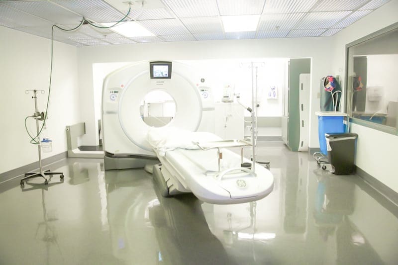
Computed Tomography (CT)
A CT scanner combines X-rays with advanced computer processing technology to create accurate detailed images of your internal structures and organs. CT exams are quick and comfortable. You will be asked to lie still on a table as it gently moves you through the scanner. Be sure to inform your physician or the technologist if you have any allergies or believe you are pregnant. CT scans allow doctors to see images of your internal organs and structures in great detail from a variety of angles.
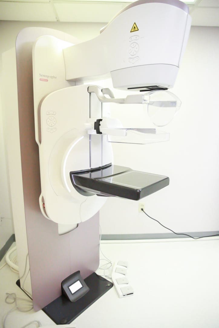
Mammography
A mammogram is an x-ray test that produces an image of the inner breast tissue on film. This technique, called mammography, is used to visualize normal and abnormal structures within the breasts. Mammography, therefore, can help in identifying cysts, calcifications, and tumors within the breast. It is currently the most effective way to detect early breast cancer. Breast self-examination (BSE) on a monthly basis and examination by a doctor are still important, but physical examinations typically find breast cancers when they are much larger than those detected by mammography.
General X-Ray
X-rays are a form of electromagnetic radiation just like visible light. They can be emitted by specially designed machines which create photons (individual X-ray “particles”) with high energies which can pass through the body and be detected by X-ray sensitive film. Structures that are dense (such as bone) will block most of the photons, and will appear white on developed film.
Nuclear Medicine
Nuclear medicine is a medical specialty that uses safe, painless, and cost-effective techniques both to image the body and treat disease. Nuclear medicine imaging is unique in that it documents organ function and structure, in contrast to diagnostic radiology, which is based upon anatomy. It is a way to gather medical information that may otherwise be unavailable, require surgery, or necessitate more expensive diagnostic tests.
FAQ MRI
–What should I expect during my MRI?
MRI is along narrow tube. You lie down on a moveable table into the opening of the tube. You will be given earplugs due to the internal parts of the magnet producing repetitive tapping, thumping, and other loud noises.
–What is the difference between MRI and CT?
MRI uses a strong magnetic field and radio waves. CT uses ionizing radiation. MRI is useful for imaging soft tissues. CT is preferred for bone and detailed images of the chest, abdomen, and pelvis.
-Is MRI safe?
Yes. No harmful radiation is involved, and the scan is pain free.
-Will I receive any injections?
Some patients may require a contrast injection through an IV to help enhance the imaging. Gadolinium is used as the contrast agent.
-How long does an MRI take?
The duration varies depending on the body part being imaged. On average exams take about 20-30 minutes; however, some can take over an hour.
–Does MRI have a size and weight limit?
Yes. The weight limit for our MRI is 350 pounds and the diameter is about 22 inches.
-How do I prepare for my MRI?
Because MRI has a very strong magnetic field, you will be asked to remove all metal and magnetic objects before entering the room. This may include: jewelry, clothing, phones, keys, wallets, hearing aids, eyeglasses, hair accessories.
Eat as you would normally and continue to take your medicines unless told otherwise.
-Other considerations:
It is important to inform your provider if you have any metal implants, devices, or foreign body. If you have anxiety or claustrophobia, let your healthcare provider know in advance. They may be able to provide options to make you feel more comfortable.
FAQ CT
-How should I prepare for my CT scan?
Eat and drink as you normally would, unless told otherwise. You may be asked to remove clothing and jewelry depending on the area being scanned. You may be asked to put on a gown. You will lie on a flat long moveable table. The table will move in and out of a donut shaped scanner. It is important to be still as movement can blur the images. Depending on your scan, you may be told to breathe in, breathe out, or to hold your breath. When the test is over, the table will move out of the scanner.
-How long will my test take?
Depending on the type of CT you are having, it can take 5-30 minutes. Those needing labs drawn prior or having to drink contrast may take longer.
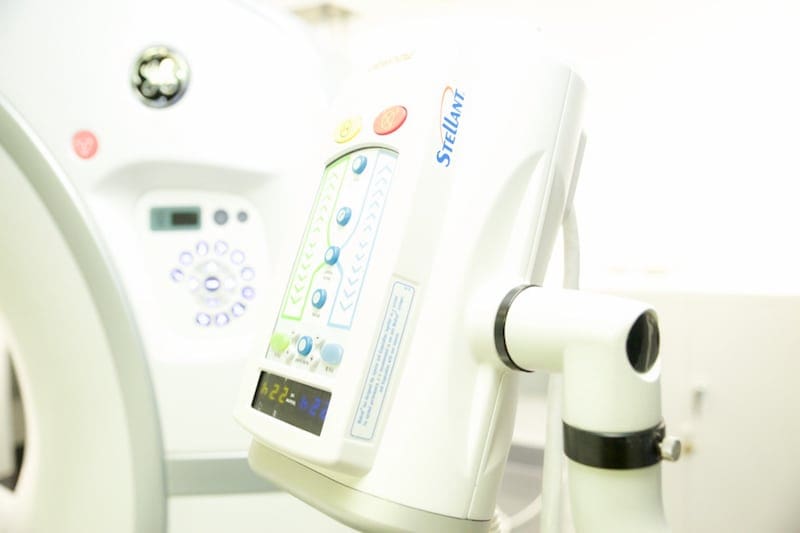
-Will I need contrast?
Depending on your healthcare provider’s order, you may need contrast. Oral contrast fills the colon and small bowel for better visualization. It can take up to two hours for oral contrast instructions. Contrast IV enhances all vascular structures on the image and characterizes any pathology for better diagnosis.
Other considerations:
If you are allergic to iodine or CT contrast, please let your provider and the technologist know.
FAQ Nuclear Medicine
-What is nuclear medicine?
Nuclear medicine is used in diagnosing and treating certain illnesses. They use radioactive materials, called radiopharmaceuticals, that can be injected through an IV, swallowed, or inhaled. A camera that detects radiation is placed over the body to collect images. These images show how the radiopharmaceutical acts in an organ or tissue.
-How long will my test take?
Depending on your exam, the test can take up to 3 hours.
-Are there any instructions I should know about?
Depending on the test, you may have specific instructions that must be followed. Please call the radiology department if you are unsure.
FAQ Mammography
-Why do mammograms squeeze my breast?
During a mammogram, your breast is compressed to decrease the breast thickness to make abnormalities more visible. The compression only lasts for a few seconds.
-What is a 3D mammogram?
3D mammography allows breast tissue to be viewed in individual segments. 3D deters 41% more invasive breast cancers and reduces false positives by 40%.
-What is the difference between a screening mammogram and a diagnostic mammogram?
Screening mammograms are used to detect early breast cancer in women with no symptoms. A diagnostic mammogram is used to evaluate abnormal symptoms such as a breast lump, abnormal discharge, inverted nipple, and breast pain. A diagnostic mammogram can target the area for a better diagnosis.
-Do I need a referral to get a mammogram?
You can schedule a screening mammogram without a referral if you’re 40 years of age and have a primary healthcare provider.
A diagnostic mammogram however does require a referral. Call your healthcare provider for your diagnostic mammogram referral.
FAQ Ultrasound
-What are the different kinds of ultrasound?
Ultrasound is used for a wide range of imaging such as pregnancy, abdominal, breast, pelvic, thyroids, hearts, veins and arteries.
-How do I prepare for my ultrasound?
Preparation will depend on the type of ultrasound you are having. For a pelvic or pregnancy ultrasound, you may need to fill your bladder. For an abdominal ultrasound you may need to fast.
-Are ultrasounds safe?
Yes. Research has shown ultrasound to be safe with no harmful side effects.